Cardiovascular and Interventional Radiological Society of Europe
CIRSE insider - NEW - animated
PublicationsCIRSE InsiderCIRSE 2026 Best Scientific Paper Award: a Q&A with the winner

CIRSE 2026 Best Scientific Paper Award: a Q&A with the winner

October 5, 2026

Christian Roemer, winner of the CIRSE 2026 Best Scientific Paper Award, shares the story behind his and his team’s research paper, ” AI-derived fully automated 3D body composition predicts outcome after transjugular intrahepatic portosystemic shunt placement“, from developing the study to presenting his findings at his first CIRSE Annual Congress.


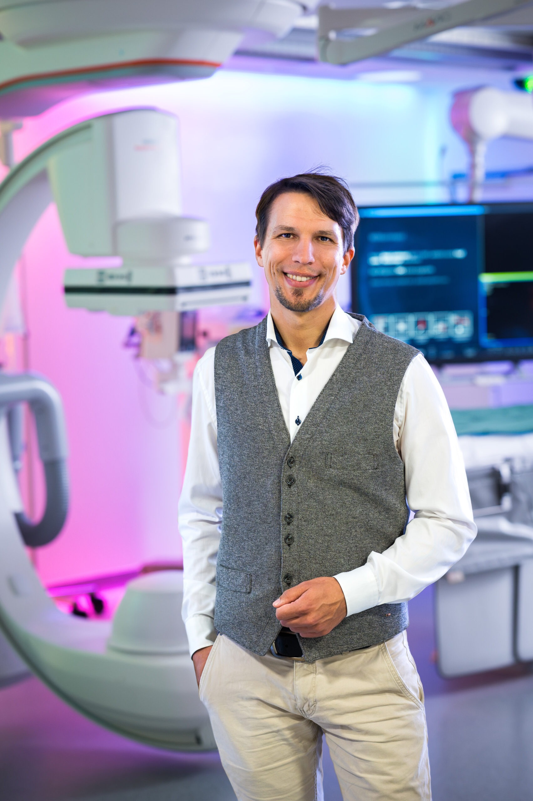
Christian Roemer

Mr. Roemer, congratulations on receiving this year’s Best Scientific Paper Award! Can you tell us a little bit about your academic background and your institution? How many TIPS procedures are performed annually?

Roemer: Thank you very much. It was a pleasure presenting our work at CIRSE, and receiving the award was a great honor for the entire team.

I studied computer science at TU Dortmund, focusing on data science, and I worked as a data scientist for several years before joining the Clinic for Radiology at University Hospital Muenster. Currently, I am a research associate in RACOON, the Radiological Cooperative Network of the German Network University Medicine (NUM), and I am completing my doctorate in medical sciences.

The University Hospital of Muenster is a high-volume centre with extensive experience treating patients with portal hypertension. The Clinic of Radiology and the Department of Medicine B (Gastroenterology and Hepatology) work together to determine TIPS indications. Our experienced IRs and gastroenterologists routinely perform the procedure together, which is central to our TIPS programme. Around 120 to 130 TIPS procedures, including emergency TIPS implantation, are performed annually. Each patient receives a standardized pre-procedural CT scan, and we document follow-ups, including shunt revisions. This combination of imaging and long-term outcome data made our study possible.

What prompted you to investigate AI-derived 3D body composition as a prognostic tool in patients undergoing TIPS placement?

Roemer: Our clinic has a long-standing collaboration with the West German Cancer Center at University Medicine Essen. Together, we have conducted several two-centre studies on body composition markers derived automatically from routine staging CTs in oncology cohorts. Across these groups, muscle volume and fat compartments were consistently related to outcomes, in some cases surprisingly strong, and independent of BMI. The idea is that the images we already acquire contain prognostic information that we do not currently use.

TIPS was a natural next application. Sarcopenia and malnutrition are common problems in cirrhosis. Every TIPS patient has a recent CT scan, and my mentors in radiology are directly involved in the procedures. We had a standardized imaging archive and well-documented endpoints, including shunt revision. We also had a fully automated pipeline that could process all of this information without manual contouring. The question was whether the same markers that predict survival in cancer patients also provide information about survival and shunt function after TIPS.

What was the process like submitting the paper to CIRSE 2026?

Roemer: With 22 candidate markers and three endpoints, the word limit forced us to narrow the focus of the abstract. We identified two key findings: the myosteatotic fat index for survival and liver volume for stent failure. We collaborated closely with our clinical partners on the abstract to ensure the imaging methods and clinical endpoints were sound.

Myosteatotic fat index (MFI) emerged as an independent predictor of mortality, whereas conventional L3 single-slice measurements did not predict overall or transplant-free survival. Why do you think volumetric 3D analysis provides more prognostic information in this patient population?

Roemer: In our cohort, conventional L3 indices, such as skeletal muscle index, did not predict survival. However, the volumetric MFI did. The MFI describes how much of a patient’s fat has accumulated inside and between the muscles rather than elsewhere in the body, which is information that muscle area alone does not capture. A post hoc analysis of the MFI on a single L3 slice also predicted survival, albeit with a weaker effect. In our oncology cohorts, the volumetric indices were clearly superior and retained their significance after adjusting for L3 indices.

Volumetric analysis provides additional benefits over a single slice. Liver volume, our only predictor of stent failure, has no single-slice counterpart. Cardiac and thoracic fat compartments, as well as bone, are not captured at L3 at all. There is also no single landmark to pick and no time-consuming manual contouring. The pipeline runs on the whole scan and returns all of these values for every patient.

What does the association between a higher MFI and mortality tell us about the importance of muscle quality and body composition in patients undergoing TIPS?

Roemer: In our data, muscle quantity did not predict survival, but muscle quality did. Patients with the same muscle volume and liver scores can differ substantially in the amount of fat infiltrating their muscle. Per standard deviation, this was associated with a 37% higher risk of death, independent of age, sex, and MELD.

Myosteatosis reflects the patient’s metabolic state. It is linked to insulin resistance, chronic inflammation, and reduced muscle function, all of which are common in advanced cirrhosis but not necessarily captured by liver-specific scores. While MELD describes the liver, body composition describes the patient’s reserve. This reserve may influence how well a patient tolerates and benefits from a shunt.

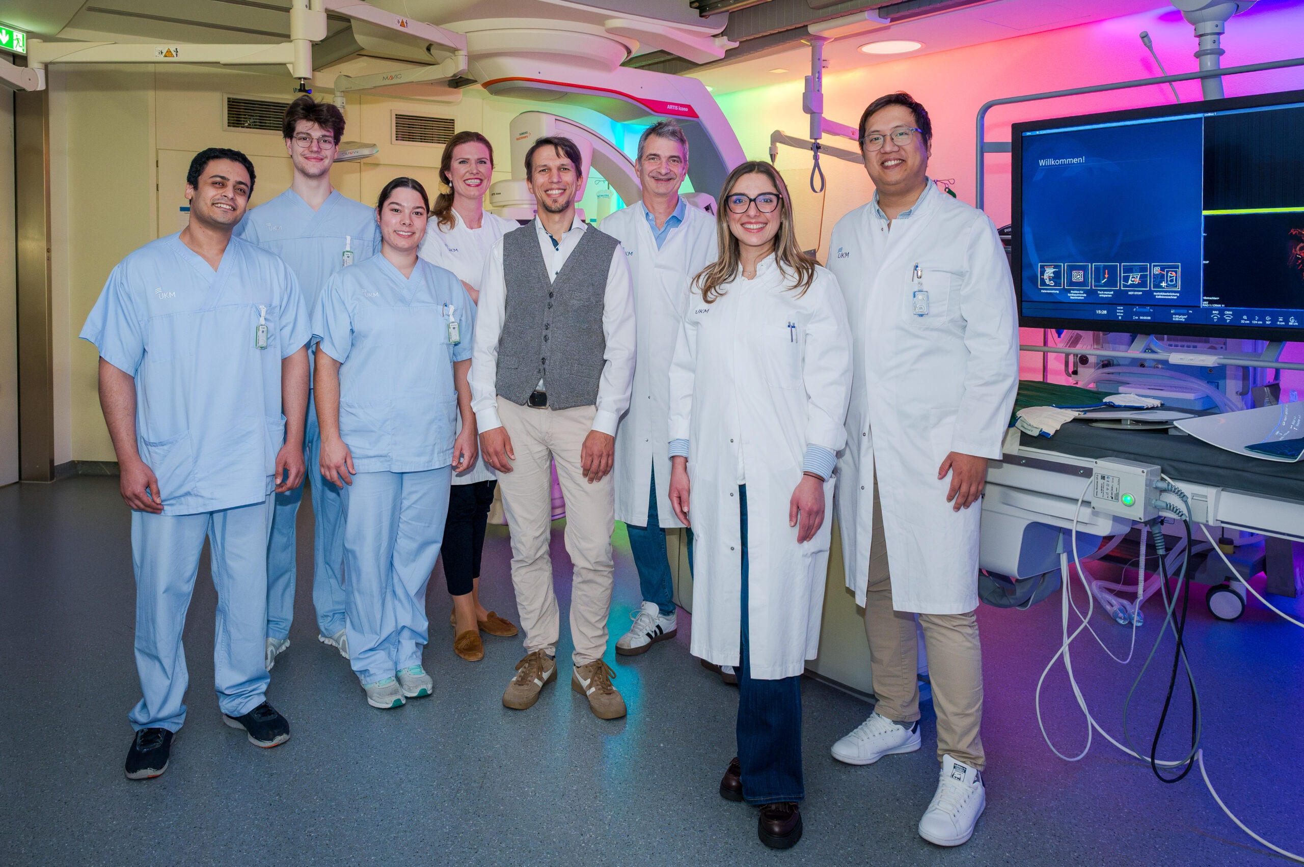
The University Hospital of Muenster, Clinic of Radiology team. From left to right: Mahmoud Younis, Florian Thomes, Susana Afonso, Gesa Poehler, Christian Roemer, Michael Koehler, Nabila Gala Nacul Mora, and Michael Praktiknjo (Department of Medicine B, Gastroenterology and Hepatology).

Out of the six markers, liver volume was the only independent predictor of stent failure. Was this finding expected? What might explain the relationship between liver volume and stent failure?

Roemer: This was unexpected. We had hypothesized that stent failure would be related to muscle or fat compartments, but it was not. Liver volume was the only marker associated with time to the first revision for undershunting.

The mechanism behind this finding is still unclear. Hypotheses raised after the presentation and within our team point toward metabolic status and comorbidities. A larger liver in cirrhosis may reflect disease stage, etiology, steatosis, or diabetes. All of these could plausibly affect shunt patency, but for now they remain hypotheses.

Because the analysis can be computed automatically from CT imaging, how could this type of AI-based assessment potentially be incorporated into clinical practice? Do you believe it could assist with patient selection, risk stratification, or follow-up after TIPS?

Roemer: The analysis requires no additional imaging, contrast, or patient time. The pre-TIPS CT scan is already available, the segmentation runs automatically, and the result is a set of numbers that can be included in the radiology report alongside the standard findings.

The most immediate use is risk stratification. The MFI and liver volume can be reported for every pre-TIPS CT, and after validation, they can be combined with the MELD score to create a risk score. For patients with a high MFI, this could prompt more intensive nutritional and metabolic optimization around the procedure. For patients with a large liver volume, closer shunt surveillance after TIPS could be considered because their probability of requiring an undershunting revision is higher.

Whether these markers should influence patient selection is uncertain. Our data show associations; whether changing management on the basis of these numbers improves outcomes is a different question, which requires external validation and, ideally, prospective data. For now, we can say that this information is available at no additional cost and that we are not yet using it.

What was your experience presenting at CIRSE 2026?

Roemer: This was my first CIRSE, and it was a great experience to present in a scientific session with both junior researchers and highly experienced IRs. The discussion after my presentation was lively and useful. Several colleagues suggested metabolic and comorbidity-related explanations for the liver volume finding. This feedback has already influenced our plans for the next analyses.

Do you have any plans to build on this research or have any upcoming research projects that you’re looking forward to?

Roemer: The results should be validated in an independent TIPS cohort and, ideally, with a prospective registry that routinely records body composition. To understand the liver volume results, we need to adjust for stent type and consider comorbidities such as diabetes and the etiology of cirrhosis. We also plan to derive sex-specific cut-off values and test a combined score with MELD. Ultimately, we aim to transition body composition from a research measurement to a standard component of the pre-TIPS CT report.

I am grateful to my mentors, PD Dr. med. Gesa Poehler and PD Dr. med. Michael Koehler, for their guidance on this project, to the body composition team at University Medicine Essen for the Body and Organ Analysis (BOA) pipeline, and to Prof. Michael Praktiknjo and Nancy Farouk from the Department of Medicine B for their TIPS cohort and clinical expertise, as well as for their ongoing support.