Cardiovascular and Interventional Radiological Society of Europe
Slider
PatientsPatients general informationIR proceduresEndovascular treatment of aortic dissections

Endovascular treatment of aortic dissections

What is an aortic dissection?

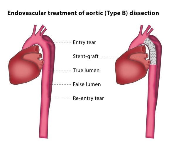
The aorta is the largest artery in your body. An aortic dissection is when the layers of the aortic wall are separated by a tear in the inner wall. The aortic wall splits into two lumens (channels): a true lumen, meaning the original lumen; and a false lumen, which is in the aortic wall. If there is high blood pressure in the false lumen, the true lumen is compressed. This decreases blood supply to the organs, leading to organ failure and restricted blood supply to the lower limbs.

There are two broad types of dissection – a tear to the artery near the heart to the point where the artery supplying the left arm takes off called Type A and a dissection beyond the origin of the artery supplying the left arm called Type B. Type A dissections need immediate surgery to repair it. Certain types of Type B dissections benefit from placement of a stent.

How does the procedure work?

The placement of a stent graft (a metallic scaffold with a surrounding fabric) is usually performed under general anaesthesia. The interventional radiologist will access an artery at the top of your leg for the procedure. They may also insert an angiography catheter through an artery in your right arm for injections of contrast material before and after the placing of the stent graft. The interventional radiologist will place a guidewire into your aortic arch and will then pass the stent graft over the wire and use this to cover the tear on the inner wall of the aorta. This protects the true lumen from the expansion of the false lumen.

If your case is critical, you may need to undergo additional stenting of these vessels to avoid restricting the blood supply.

If a stent graft is not enough to decompress the false lumen, you may be advised to have a procedure called fenestration of the intimal flap. This means that a hole will be made in the membrane between the true and false lumens to equalise the pressure between them. The interventional radiologist will create a connection between the lumens using a needle and positioning a wire through the puncture site. They will then widen the hole using a tiny balloon and may implant a stent to keep the connection open.

Why perform it?

The purpose of these procedures is to restore the true lumen as well as the blood supply to your organs and lower limbs. The procedures also stabilise the pressure and blood flow in the area and protect the aorta from widening.

What are the risks?

Depending on the location of the aortic tear, it may be necessary for the interventional radiologist to place the stent graft over the origin of an artery in your chest, which would result in a decreased blood supply to your left arm. This may lead to weakness of the arm, dizziness and ischaemic injuries to your brain. If you experience these symptoms, the artery will be treated to improve blood flow.

In some cases, it is not possible to separate the false lumen, which would make it necessary for you to undergo further treatment. Other risks include the misplacement of the stent graft and the rupture of the vein used for access, but these are rare.

Bibliography

1. Fanelli F, Dake MD. Standard of practice for the endovascular treatment of thoracic aortic aneurysms and type B dissections. Cardiovasc Intervent Radiol 2009; 32:849-860.