What is brachytherapy?
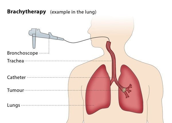
Brachytherapy is a form of therapy used to treat cancer by placing radiation inside the body.
Brachytherapy is a form of therapy used to treat cancer by placing radiation inside the body.
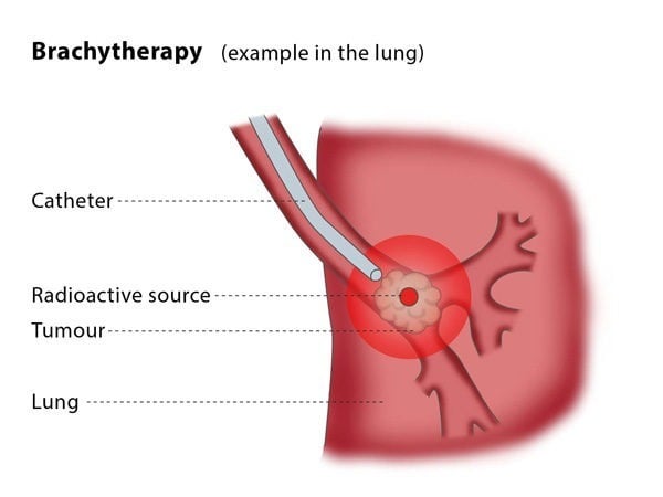
The interventional radiologist will place small metal beads or wires into the cancerous tissue, usually using radiological imaging as a guide. These emit high energy radiation, which then spreads a short distance from this source into the surrounding tissue containing the tumour, whilst limiting radiation to the non-cancerous, normal tissues that are further away. The radiation kills the tumour cells.
The procedure is performed to cure cancer or to limit its progression. It may be used alone or conjunction with other therapies including chemotherapy and surgery. It has been applied to a range of cancers including, but not limited to, prostate, cervix, liver, rectum and breast cancer.
Placing the radiation source carries the risk of bleeding and infection. There is a risk of the radiation killing normal, non-cancerous cells. You may experience pain in the area treated.
1. Collettini F, Golenia M, et al. Percutaneous computed tomography-guided high-dose-rate brachytherapy ablation of breast cancer liver metastases: initial experience with 80 lesions. J Vasc Interv Radiol 2012 23(5): 618-626.
2. Luo J, Yan Z, et al. Endovascular placement of iodine-125 seed strand and stent combined with chemoembolization for treatment of hepatocellular carcinoma with tumor thrombus in main portal vein. J Vasc Interv Radiol 2011 22(4): 479-489.