Cardiovascular and Interventional Radiological Society of Europe
Slider

Bone augmentation

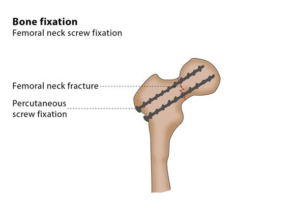
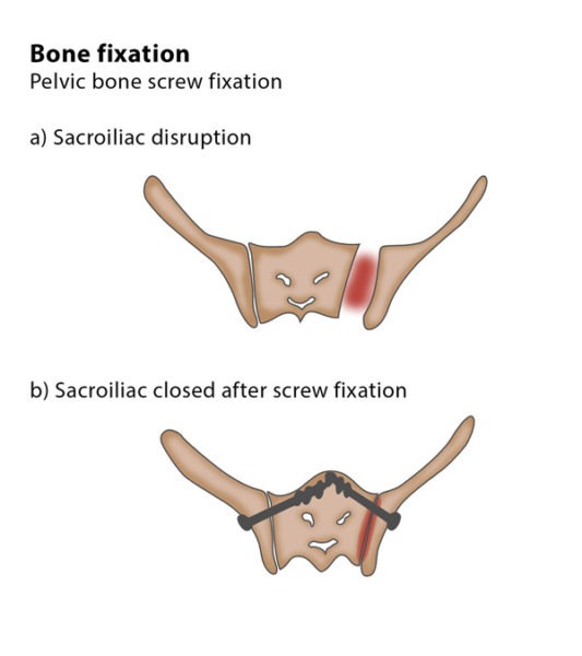
What is bone augmentation?

Bone augmentation techniques aim to stabilise a weakened or a fractured bone. These minimally invasive techniques include injection of bone cement designed for this purpose, the insertion of metallic rods or screws, or a combination of both techniques, depending on the location and on the type of fracture.

How does the procedure work?

The interventional radiologist will use image guidance to precisely insert the devices used to reinforce a weakened or fractured bone.

Why perform it?

Percutaneous image-guided bone fixation is performed to stabilise a weakened bone. The procedure is a minimally invasive alternative to conventional surgery and it may be recommended to treat a range of conditions, including bone metastases, osteoporotic fractures or fractures caused by trauma.

What are the risks?

Placing needles inside bone carries the risk of bleeding and infection. In rare cases, the surrounding structures such as nerves or vessels are damaged. Occasionally, optimal stabilisation cannot be achieved, which can cause a delayed fracture to another bone. You are especially at risk for this if you have advanced bone cancer.

Bibliography

1. Huegli RW, Messmer P, Jacob AL, Regazzoni P, Styger S, Gross T. Delayed union of a sacral fracture: percutaneous navigated autologous cancellous bone grafting and screw fixation. Cardiovasc Intervent Radiol. 2003 Sep-Oct; 26(5):502-5.
2. Trumm CG, Rubenbauer B, Piltz S, Reiser MF, Hoffmann RT. Screw placement and osteoplasty under computed tomographic-fluoroscopic guidance in a case of advanced metastatic destruction of the iliosacral joint. Cardiovasc Intervent Radiol. 2011 Feb; 34 Suppl 2:S288-93.
3. Deschamps F, Farouil G, Hakime A, Teriitehau C, Barah A, de Baere T. Percutaneous stabilization of impending pathological fracture of the proximal femur. Cardiovasc Intervent Radiol. 2012 Dec; 35(6):1428-32.