Cardiovascular and Interventional Radiological Society of Europe
Slider
PublicationsCIRSE InsiderCIRSE Fellowship Grant Programme: Dr. Kevin Fung

CIRSE Fellowship Grant Programme: Dr. Kevin Fung

March 8, 2024

The CIRSE Fellowship Grant Programme (FGP) allows CIRSE members to apply for a grant to fund a one-month stay at a European centre outside their country of residence, where they can train or experience new procedures. With this programme, the CIRSE Foundation actively supports international mobility of IRs and the growth of IR across Europe.


By Dr. Kevin Kin Fen Fung

It is a such a great honour to be a recipient of the 2024 CIRSE Fellowship Grant to further my interventional oncology training for a month in December 2023 at the Department of Interventional Radiology at the University Hospital Strasbourg under Professor Afshin Gangi and his amazing team.

I am a paediatric interventional radiologist (IR) and the lead of the IR team at Hong Kong Children’s Hospital. Our hospital takes care of all the paediatric oncology patients in Hong Kong, a metropolitan city in East Asia with a population of 7 million people. We face many challenging oncological cases such as paediatric liver tumours, sarcomas, desmoid tumours and lung metastases. With our paediatric oncologists, surgeons and IRs, we take a multidisciplinary (MDT) approach to manage these complex patients in a child-centred manner. Currently at our centre, the IRs are taking an increasing role in the patient journey, participating in joint clinics, MDT meetings and consultations. When children with suspected cancer present to our centre, the IRs help expedite the diagnosis by performing image-guided biopsy and provide image-guided long term central venous access. Most of the children receive these essential procedures within three days of presentation to our hospital, and we are able to initiate chemotherapy within one week. For tumours resistant to chemotherapy or relapse after surgery, our IR team have started to offer ablation and chemo-embolization for oncologic control.

Group photo with Prof. Gangi, Dr. Bertucci, Dr. Caudrelier, Pol and Guillaume
Group photo with the IR radiographers and nurses

Professor Gangi and his team are world-renowned for state-of-the-art interventions in the field of interventional oncology, particularly in ablation for a wide variety of benign and malignant tumours. His department is also IASIOS-accredited, with aim of delivering high quality patient-oriented interventional oncology care. This one-month observership has provided me with a great opportunity to learn the following from Professor Gangi and his team:

  • Setup, workflow, clinical indication and technical aspects of a wide variety of innovative ablation therapies for benign and malignant tumours, including cryoablation, interstitial laser ablation, microwave ablation, irreversible electropolation and electro-chemotherapy
  • Setup and workflow for Angio-CT and Interventional MR, and how these modalities can be best tailored to different IR procedures
  • Joint clinical management of desmoid patients in a multidisciplinary setting with oncologists to provide the best-tailored plan for the patient
  • Setup of a well-run IR clinic and IR ward
  • The process of IASIOS accreditation and the practical aspects of delivering a highly effective clinical IO service

Professor Gangi’s Department manages a very high volume of patients every day. For the non-vascular interventional section alone, the Angio-CT and single plane angiography run from 8:30 in the morning till 6 to 7pm in the evening, and there is interventional MR on most mornings. The ablation treatments (known as “les grandes gestes” – literally “big procedures”) usually take place in the morning, with the anaesthetist preparing the patient at around 8:00am. In the afternoon, the list is busy with image-guided biopsies and joint injections. There is an IR clinic almost every day where the patients are evaluated by an interventional radiologist and the treatment options are discussed with them. Professor Gangi, his co-chair Prof. Garnon, Dr. Cazzato (probably better known as “Gigi”), Dr. Caudrelier, Dr. Koch, Dr. Pierre-Alexis, Dr. Bertucci and Dr. Weiss, together with a dedicated team of radiographic technicians and nurses, were all great teachers and enthusiastic to share their knowledge and answer any questions I had. I felt very welcome as part of the team.

View of University Hospital Strasbourg
Selfie with Prof. Gangi
The setup at the Angio-CT suite

On the technical side, I was most impressed with the ingenious use of hydrodissection and carbo-dissection for the protection of critical structures during ablations. There was one patient with three hepatocellular carcinomas (HCC) located at challenging locations, necessitating hydrodissection of the gallbladder (for the subcapsular segment 5 HCC), bare area (for segment 4a HCC) and hepatic hilum (for centrally located HCC). It is amazing how a 22G needle with continuous saline infusion can make all the difference and expand the realm of ablative treatment to tumours located in difficult locations. Another case which really wowed me was using hydrodissection to protect the laryngeal and pharyngeal structures during cyroablation of a cervical nodal metastasis. For this patient, Prof. Garnon had to put his finger into the patient’s mouth to ensure the buccal mucosa was not frozen!

The use of an Angio-CT machine also greatly enhances the workflow and efficiency of ablations. It has the capability of planning a double-oblique path which makes performing cyroablation or irreversible electroporation easier as these modalities often require the insertion of multiple probes in a parallel fashion. For patient requiring bone consolidation after ablation of osseous metastasis, the fluoroscopic component of the Angio-CT allows more accurate and safer injection of cement under continuous monitoring.

Interventional MR was another innovative technique which I believe would have great utility in children, given its lack of radiation and real-time visualisation of iceballs for cyroablation. Prof. Gangi and his team have thoroughly deliberated the logistics aspects of the interventional MR setup, which is essential for a smooth and safe procedure. The argon and helium required for cyroablation are installed as wall outlets so there is no issue of running out of gas or placing multiple gas canisters outside the MR suite. They have also tethered an ultrasound (US) machine within the 5-gauss line in the MR suite to allow for quick US-guided needle positioning whilst ensuring the machine would not be inadvertently drawn to the magnet. A mobile MR-compatible TV monitor was installed in the MR suite to provide real time image guidance during needle manipulation. Appropriate hearing protection was also provided to all the personnel in the room as it gets very loud during the real-time sequence acquisition. These are all important aspects which I need to consider when setting up an interventional MR service back in Hong Kong.

Photo with Prof. Julien Garnon, Ms. Mardis Karlsdottir (Chief operating officer from IASIOS) and Robert Hack (Videographer from the CIRSE team)
The setup at the Interventional MR suite
The dedicated storage for argon gas

I am very impressed by how well IR has been integrated into the patient journey here at the University Hospital Strasbourg. After receiving referrals or consultations from other specialties, interventional radiologists attend to the patients directly to explain and discuss treatment options in the clinic or in the ward. IRs also have rights to admit patients under their care as in-patients. After the procedure, if the patient does well, he or she can be discharged back home directly. According to the clinical condition, the IRs determine the necessary imaging and clinical follow up schedule. Putting myself in the patient’s shoes, this seamless workflow allows for a better understanding of the treatment plan and continuity of care. For patients with more complex oncological conditions such as desmoid fibromatosis, I had the pleasure of sitting on the joint clinic where interventional radiologists and oncologists can assess the patient together and offer a tailored treatment plan. In cases unresponsive to chemotherapy or tyrosine kinase inhibitors, cyroablation can be performed as a definitive treatment to shrink down the tumour or as symptomatic control for pain relief. It is very uplifting to see the joy some of these patients expressed during their follow up clinic visit.

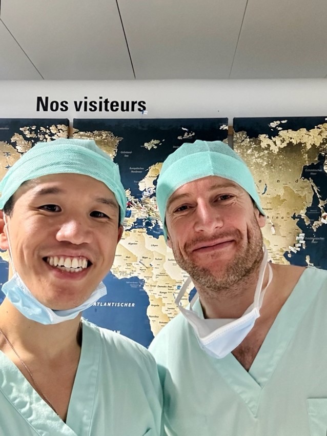
Quality and safety are very important aspects in clinical care and it is encouraging to see CIRSE taking the lead to help standardise quality assurance in interventional oncology through the International Accreditation System for Interventional Oncology Service (IASIOS). Professor Gangi’s Department is the second in the world to be accredited by IASIOS. I was able to observe in first-hand how IASIOS has optimized the daily practice of IR by streamlining processes, reducing delays, and ensuring accurate patient documentation. It was also opportune that Miss Mardis Karlsdottir, the Chief Operating Officer of IASIOS, visited Professor Gangi’s Department during my time there. I had the chance to gain a deeper understanding of the accreditation process and the benefits of IASIOS, including commitment to high quality clinical IO service and promotion of patient safety.

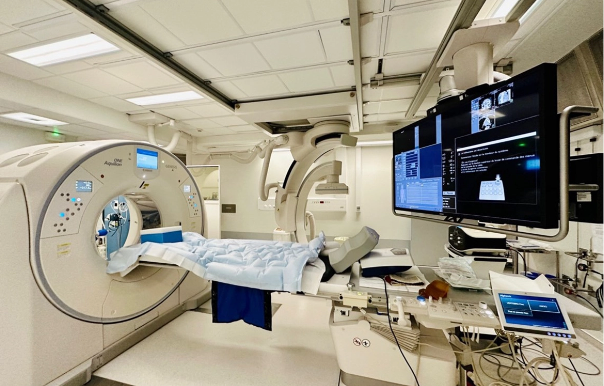
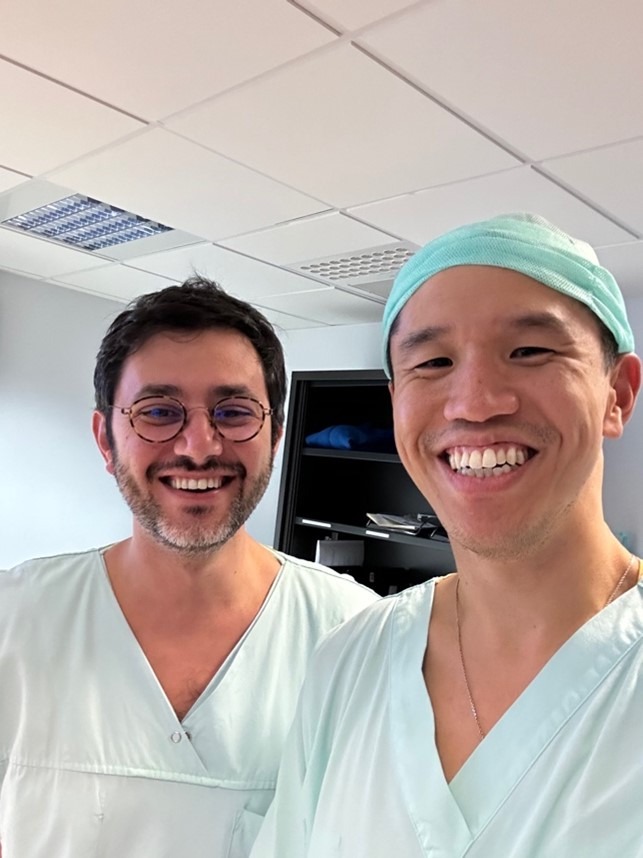
In addition to patient care, Professor Gangi and his team are also active researchers and pioneers in innovative therapies. Together with Dr. Cazzato, I had the chance to review MR-guided transurethral ablation (TULSA) as a novel minimally invasive treatment for patients with localised prostatic cancer. Conventional treatments for these men are radical prostatectomy and external beam radiotherapy, which both carry significant treatment-related genitourinary functional complications. MR-guided TULSA utilises real-time MR imaging and thermometry to ablate prostate tissue and has been shown to improve functional outcomes whilst maintaining satisfactory oncologic control. I am very pleased that our review article on MR-guided TULSA has been accepted by the CVIR Journal and will be published soon.

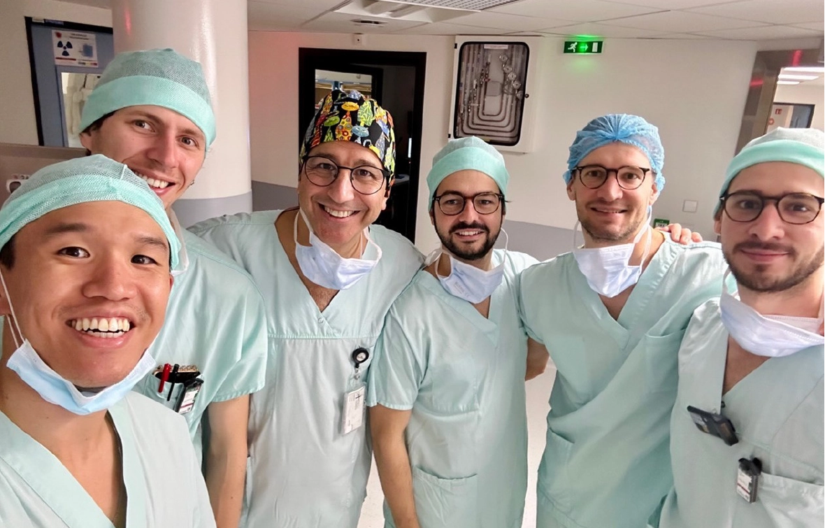
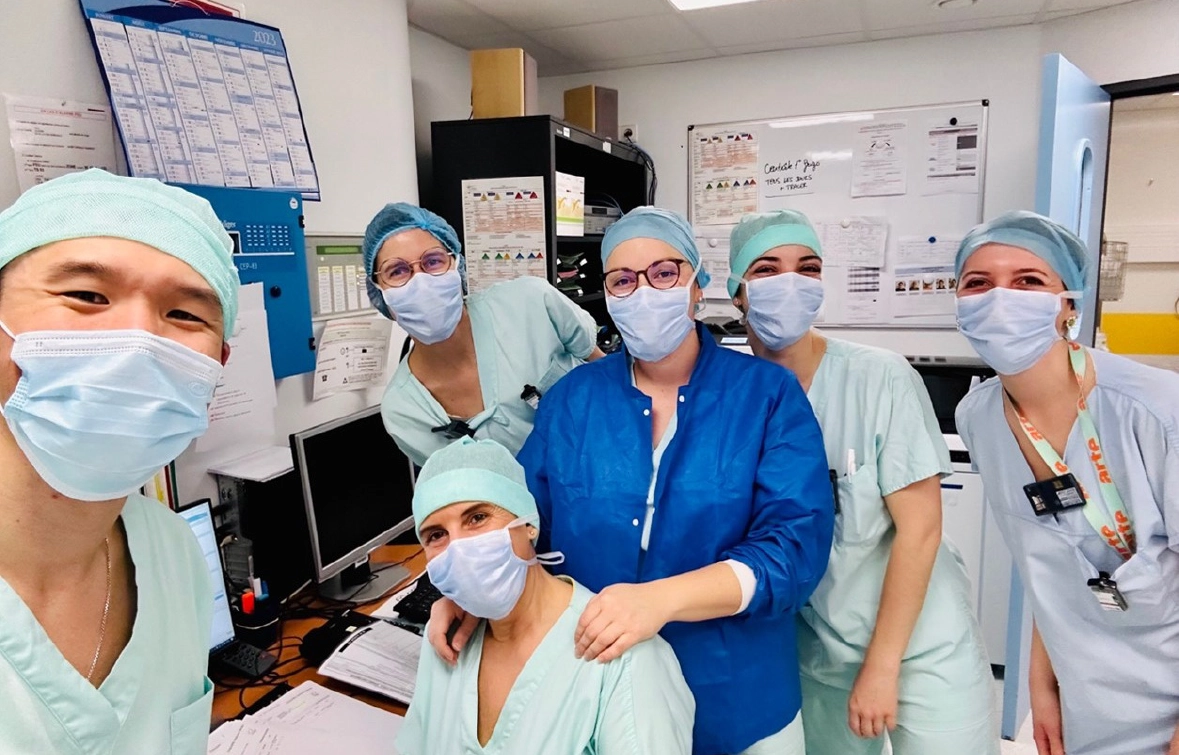
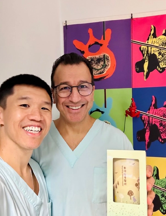
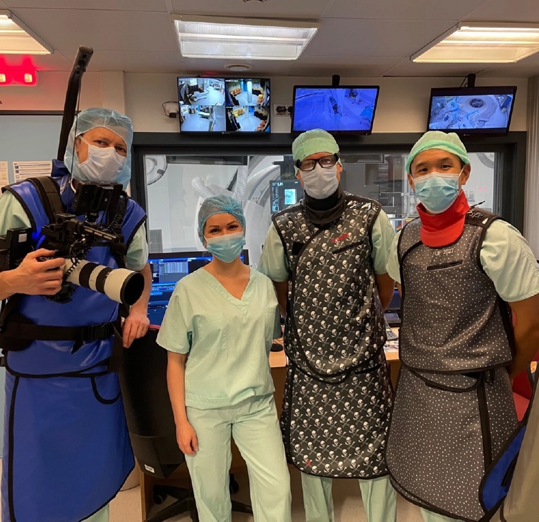
I have gained so much during my one-month time attaching at Professor Gangi’s Department, not only in terms of IR-related knowledge and insights but also many friendships. Professor Gangi is a great mentor, clinician and scientist – warm, kind, caring and innovative. He is a very busy man yet he manages to find time to look after the Fellows and take care of his patients amongst many other duties. Everyone in his team is full of energy, dedicated and friendly. Despite the little French I know, they were kind enough to invite me to their departmental Christmas party which was an absolute blast. I would highly recommend any IRs with interest in developing interventional oncology services to visit Professor Gangi and his team to experience clinical IO at its best!

Selfie with Prof. Garnon
Selfie with Dr. Cazzato
Fun moments at the Christmas party (Dr. Bertucci’s alternative career is a DJ!