Cardiovascular and Interventional Radiological Society of Europe
Slider

Jejunostomy

What is percutaneous image-guided jejunostomy?

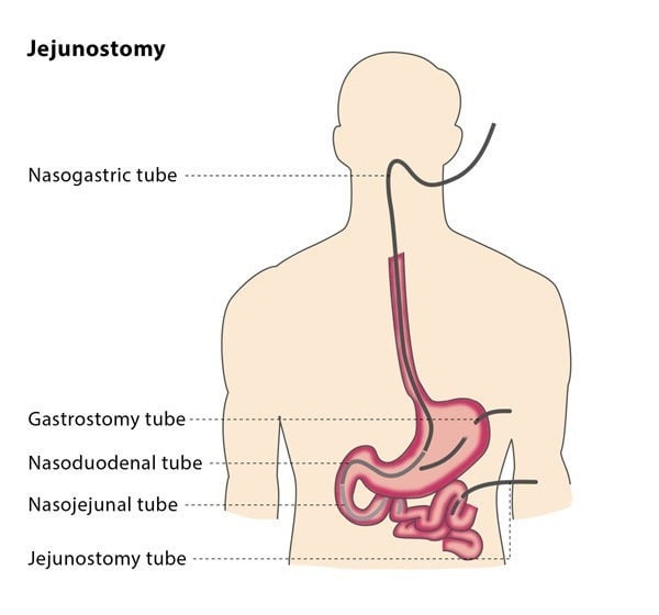
If you have been unable to eat for over seven days, you may need to receive nutrients intravenously (called parenteral feeding) or via a tube into the stomach or small bowel (called enteral feeding). There are a number of ways that enteral feeding can be carried out, such as placing a tube through the nose and into the stomach. This is called a nasogastric or nasoenteric feeding tube, and is the preferred option if your anticipated need is less than 30 days.

However, nasoenteric tubes are not suitable for use longer than 30 days, as they can cause considerable discomfort and complications such as inflamed sinuses. If your need is anticipated to be for longer than 30 days, a better option for you is direct enteral access. This involves placing a tube directly into your stomach or bowel. This used to require surgery, but minimally invasive techniques are now available as an alternative, such as percutaneous image-guided jejunostomy.

During a percutaneous image-guided jejunostomy, an interventional radiologist will place a tube directly through the abdominal wall and into part of your small intestine called the jejunum, providing a way for nutrients to enter your body.

How does the procedure work?

The procedure is carried out using fluoroscopic guidance. The interventional radiologist will first inflate your stomach using a nasogastric tube. This will aid the fluoroscopy, the required puncture and enlargement of the tract. In rare cases, nasogastric access may not be possible, so the stomach is inflated using a needle introduced into the stomach, under image guidance.

To minimise the risk of puncturing the colon, the interventional radiologist may administer a contrast liquid into your colon the day before the jejunostomy procedure, to help visualise the colon.

During the jejunostomy procedure, the interventional radiologist will puncture the skin where the tube will be inserted, and then direct the needle under image guidance to the small intestine. The needle may be attached to an anchor, which the interventional radiologist will direct into the jejunum using a guidewire. To ensure there is enough space for the tube, the tract will be expanded using dilators or tiny balloons, which the interventional radiologist will insert using a separate guidewire.

The interventional radiologist will then insert a jejunostomy tube over the guidewire, using fluoroscopy to confirm its position. Once it has been confirmed that the tube is correctly placed, the interventional radiologist will remove the guidewires and secure the tube to the skin using anchors.

Why perform it?

There are a number of reasons why a jejunostomy tube may be beneficial for you. The tubes may be used in infants and children who have issues with swallowing due to surgery. They may also be used in patients who are unable to swallow as a result of central nervous system disorders, patients who require feeding supplementation or special diets or to administer frequent doses of multiple medications. Patients who are chronically ill or neurologically impaired may require a tube indefinitely.

Jejunostomy is an alternative for patients who may not be able to have a feeding tube in their stomach.

What are the risks?

It is very common for air or gas to be present in the abdominal cavity as well as bruising. However, these usually have no adverse clinical effect.

Minor complications that you may experience include incorrect tube placement or tube movement, granulation tissue formation (skin that is red and sore as it heals) and minor bleeding from the site. Some patients develop a condition called intussusception, when part of the intestine folds onto another section of the intestine, causing bowel obstruction. Another possible complication is local infection.

Other procedure-related complications include damage to the abdominal or gastric walls caused by decreased blood supply and leakage from the catheter. Major complications are rare, but include perforation of the small intestine and inflammation of the lungs or airways caused by inhaling food or other substances.

Bibliography

1. Olson DL, Krubsack AJ, Stewart ET “Percutaneous enteral alimentation: gastrostomy versus gastrojejunostomy,” Radiology 1993.
2. Friedman JN, Ahmed S, Connolly B, Chait P, Mahant S, “Complications associated with image-guided gastrostomy and gastrojejunostomy tubes in children,” Pediatrics. 2004 Aug; 114(2):458-61.