What is an arteriovenous malformation (AVM)?
Arteriovenous malformation (AVM) are a tangle of blood vessels with abnormal communications between arteries and veins.
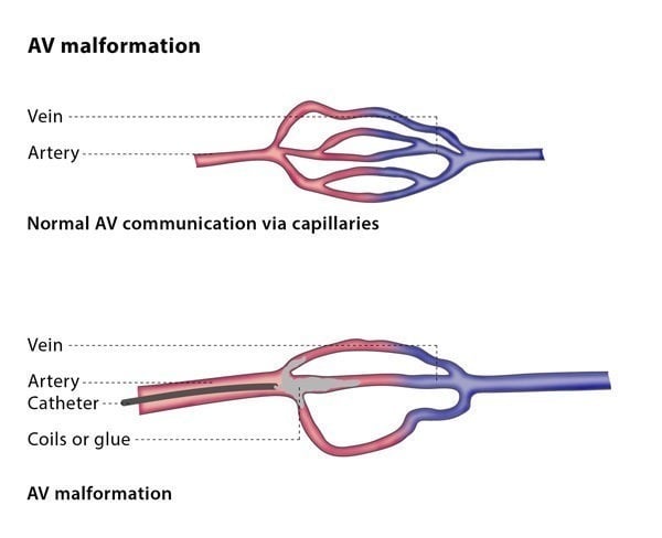
Normally, high pressure arteries are connected to low pressure veins via a network of tiny blood vessels called capillaries. Normal tissues obtain its nutrients and oxygen supply from this capillary network.
In AVMs, abnormal connections are formed between arteries and veins. These connections often skip the normal capillary bed and result in direct communication between arteries and veins. This means that normal tissues in that part of the body would be starved of oxygen and nutrients. With time, this may lead to pain and fragility in that part of the body. In addition, AVMs may grow in size over time due to rapid blood flow. This can cause reduced function of the involved organ or body part. Finally, the rapid blood flow through an AVM means that the heart needs to work extra hard to pump more blood around the body.
AVMs can occur in any part of the body, from head to toe. Different organ functions may be affected depending on its location. However, most AVMs are small, may not cause any problems and are often discovered by chance.